






Focal Microwave Ablation of the Prostate: Targeted Treatment of Prostate Cancer Aimed at Preserving Healthy Tissue
What Is Focal Microwave Ablation of the Prostate?
Prostate cancer is one of the most common cancers affecting men. When choosing a treatment, doctors consider several factors, particularly the biological characteristics of the tumour, its extent and location, biopsy results, MRI findings, and the patient’s overall health.
One treatment option for carefully selected patients is focal microwave ablation. This is an organ-preserving approach designed to destroy a specific tumour lesion without automatically treating the entire prostate.
The procedure uses thermal energy. Microwave energy generates heat in the targeted area, causing damage to and destruction of the tumour tissue. A planned safety margin around the tumour is included in the treatment plan to ensure adequate coverage of the target area.
Unlike radical prostatectomy, focal ablation does not involve removing the prostate. Preserving part of the healthy tissue may help reduce certain treatment-related side effects. However, this does not mean that complications cannot occur or that a successful outcome can be guaranteed.
How Does MRI/Ultrasound Fusion Guidance Work?
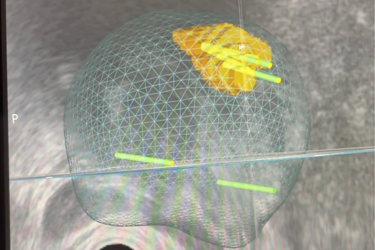
Accurately identifying the location of the tumour is particularly important in focal treatment. For this reason, the procedure may use fusion guidance, which combines information from a preoperative magnetic resonance imaging (MRI) scan with a real-time ultrasound image of the prostate.
During the procedure, an ultrasound probe is inserted into the rectum. Software then fuses the ultrasound image with the previously obtained MRI scan, which may provide more detailed information about the location of the tumour lesion. Where technically possible, the planning process may also incorporate findings from previous biopsies.
This combined image helps the physician plan the placement of the applicator needle and determine the extent of the treatment area. However, fusion guidance cannot guarantee that all tumour lesions will be identified or that the full microscopic extent of the disease will be detected.

How Is the Procedure Performed?
Focal microwave ablation is performed using a transperineal approach, meaning that the applicator is inserted through the skin of the perineum, the area between the scrotum and the anus. The exact procedure depends on the device used, the patient’s anatomy, and the individual treatment plan.
1. Preoperative assessment
The physician reviews the biopsy results, prostate MRI, PSA levels, and other relevant clinical information. The assessment also includes current medications, potential allergies, the risk of bleeding or infection, and suitability for anaesthesia.
2. Imaging and treatment planning
An ultrasound probe and MRI/ultrasound fusion are used to identify the target area. The physician plans the extent of ablation, taking into account the tumour’s location and its proximity to important anatomical structures.
3. Insertion of the applicator antenna
Under image guidance, an applicator needle or antenna is inserted through the perineum and positioned in the planned area of the prostate.
4. Treatment of the tumour lesion
Microwave energy generates heat in the target area, causing the tissue to die. Depending on the size and shape of the lesion, several applicator positions or individual energy applications may be required.
5. Post-procedure care
After the procedure, the medical team monitors the patient’s condition and urinary function. Depending on the circumstances, a temporary urinary catheter may be required. The patient receives instructions on recovery and follow-up appointments.
What Are the Benefits of Focal Treatment?
The main principle of focal ablation is to treat the known tumour area while preserving as much healthy prostate tissue as possible.
Compared with removing the entire prostate, this approach has the potential to reduce the risk of certain functional side effects, particularly urinary incontinence and erectile dysfunction. However, outcomes depend on the tumour’s location, the extent of treatment, the patient’s condition, and other factors. Neither preservation of erectile function nor urinary continence can be guaranteed.
It is also important to understand that focal treatment is not suitable for every man diagnosed with prostate cancer. Its use is determined on an individual basis following a thorough assessment of the findings and a comparison of available treatment options.
What Are the Risks and Limitations?
Prostate cancer can be multifocal, meaning that tumour lesions may be present in several areas of the prostate. Neither MRI nor biopsy is guaranteed to detect every area affected by the disease.
Following focal microwave ablation, tumour tissue may therefore persist within the treated area, at its margins, or in another part of the prostate that was not treated. Further treatment may be required in the future, including repeat ablation or another form of cancer therapy.
Possible side effects include:
- Burning or pain during urination.
- Increased urinary frequency and urgency.
- Blood in the urine or semen.
- Temporary difficulty emptying the bladder.
- Pain or discomfort in the perineal area.
- Urinary tract infection or prostatitis.
- Changes in erectile or ejaculatory function.
Less commonly, more serious complications may occur, such as significant urinary retention, severe infection, or damage to surrounding structures.
The available evidence must also be considered when making a treatment decision. The long-term oncological outcomes of microwave ablation have not been established to the same extent as those of standard radical prostatectomy or radiotherapy. Focal microwave ablation should therefore not automatically be considered equivalent to all established standard treatment options.
What Are the Alternatives for Treating Prostate Cancer?
The choice of treatment depends on the cancer’s risk category, its extent, and the patient’s individual circumstances. Available options include:
Active surveillance
Active surveillance is suitable primarily for selected patients with low-risk disease. It involves regular PSA testing, imaging examinations, and repeat biopsies when indicated.
Radical prostatectomy
This surgical procedure involves removing the prostate and seminal vesicles. For appropriately selected patients, it is a treatment with curative intent and also allows detailed histological examination of the removed tissue. Potential consequences include urinary incontinence and erectile dysfunction.
Radiotherapy
Radiotherapy uses ionising radiation to destroy cancer cells. Depending on the characteristics of the disease, it may be combined with hormone therapy.
Brachytherapy
Brachytherapy is a form of internal radiotherapy in which a radiation source is placed inside the prostate or in its immediate vicinity. It is suitable for selected patients.
Specialised centres may also offer other focal treatment methods, such as high-intensity focused ultrasound (HIFU) or cryotherapy. These methods differ in how they work, the available evidence supporting their use, and their suitability for individual cases.
Why Is Follow-Up Essential After the Procedure?
Treatment does not end with focal ablation. Long-term follow-up is an essential part of patient care and helps detect any persistence or recurrence of the disease at an early stage.
An important marker is PSA, or prostate-specific antigen. However, because part of the prostate remains intact after focal treatment and continues to produce PSA, a significant decrease in PSA levels does not, by itself, confirm that all tumour tissue has been eliminated.
Follow-up may include:
- Regular PSA testing.
- Follow-up prostate MRI.
- Repeat biopsy according to the established protocol.
- Additional examinations based on individual findings.
The specific follow-up schedule is determined by the treating centre. Some protocols involve PSA testing every three months, an initial follow-up MRI within the first few months after the procedure, and further MRI and biopsy examinations at approximately 12 months. Exact timings may vary depending on the technique used and the patient’s clinical situation.
If follow-up examinations identify persistent or new clinically significant cancer, further management is tailored to the findings. Options may include repeat focal treatment, surgery, radiotherapy, or another form of cancer therapy.
Is Focal Microwave Ablation Suitable for You?
Suitability for focal microwave ablation cannot be determined solely on the basis of a prostate cancer diagnosis. The physician must assess the histological findings from the biopsy, prostate MRI, the location and extent of the tumour, the disease risk category, the patient’s overall health, and the patient’s preferences.
Patients are advised to bring their biopsy results, prostate MRI report and available imaging files, recent PSA results, and previous urological or oncological medical records to the consultation.
The purpose of the consultation is to compare the available options, explain their potential benefits and risks, and identify the approach that best meets the patient’s individual needs.
Important: Focal microwave ablation is a targeted treatment designed to preserve as much healthy prostate tissue as possible. However, its suitability, expected benefits, and potential risks must be assessed individually. Treatment decisions should be based on specialist evaluation and an informed discussion of all available options.
At our clinic, we perform the procedure using the Koelis Trinity system.
Are you an international patient interested in focal microwave ablation of the prostate? If so, please contact us via our contact form. We will take care of all the arrangements related to the procedure. All you need to do is arrange your flights and accommodation.
Latest news


